**
Overview
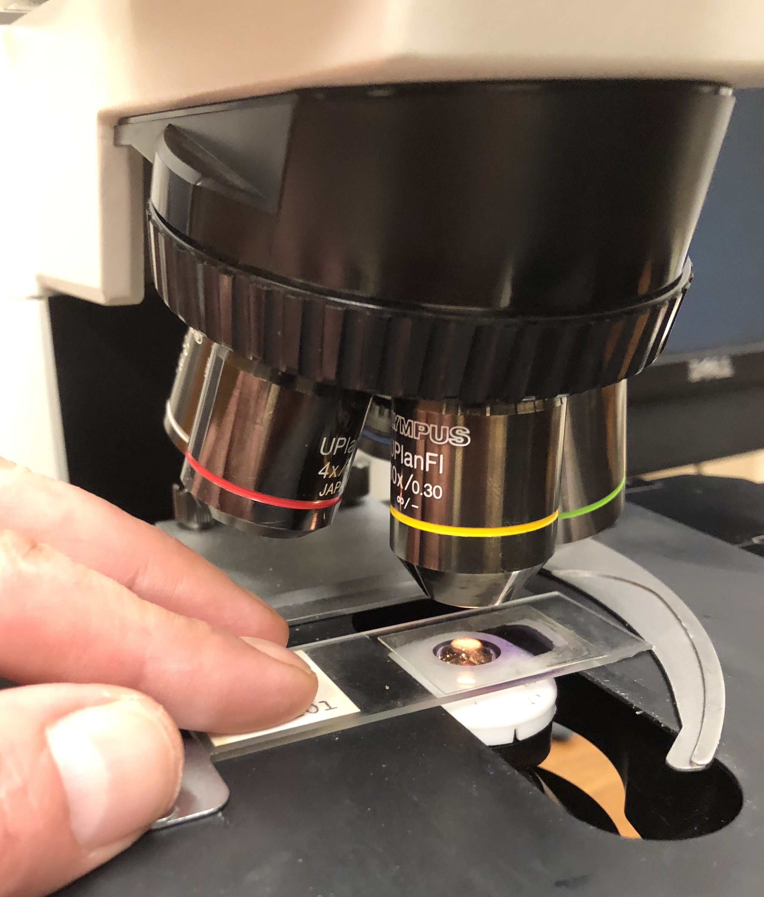
Histology—also called microscopic anatomy, microanatomy, or histoanatomy—examines tissues at magnifications ranging from 40× to 1,000×, typically using light microscopes equipped with lenses that resolve details as fine as 0.2 µm. By preparing thin sections (often 5–10 µm thick) and staining them with dyes such as hematoxylin and eosin (H&E), researchers can distinguish cell types, extracellular matrix components, and vascular networks that are invisible to the naked eye. Modern histology integrates immunohistochemistry, fluorescence microscopy, and digital image analysis, allowing scientists to map protein expression, track disease progression, and even reconstruct three‑dimensional tissue architecture from serial sections.The discipline serves as the microscopic counterpart to gross anatomy, which surveys organs and systems visible without magnification. While gross anatomy tells us where a heart sits in the thorax, histology reveals the layered arrangement of myocardial fibers, intercalated discs, and capillary networks that enable its contractile function. In medicine, histological examination of biopsy samples remains the gold standard for diagnosing cancers, inflammatory disorders, and infectious diseases, making it indispensable for both research and clinical practice.
History/Background
The roots of histology trace back to the invention of the compound microscope in the early 17th century. In 1665, Robert Hooke published Micrographia, describing “cells” in cork—a term derived from the Latin cella (small room). However, it was not until 1805 that Johann Heinrich Meckel coined the term “histology” to denote the study of tissues. The 19th century saw rapid advances: Camillo Golgi introduced the black reaction in 1873, allowing visualization of individual neurons, while Santiago Ramón y Cajal refined silver staining techniques in 1888, laying the foundation for modern neurohistology.The development of paraffin embedding in the 1890s standardized tissue processing, and the introduction of microtomes capable of cutting sections as thin as 2 µm in 1900 dramatically improved image clarity. The mid‑20th century brought immunohistochemistry (first described in 1941 by Albert Coons) and electron microscopy (commercially available by the 1970s), expanding the resolution limit to sub‑nanometer scales. In the 21st century, digital pathology and AI‑driven image analysis (emerging around 2015) have begun to automate diagnostic workflows, heralding a new era of quantitative histology.
Key Information
- Tissue Types: Epithelial, connective, muscle, and nervous tissues constitute the four primary histological categories, each with distinct cellular morphology and extracellular matrix composition. - Staining Techniques: Beyond H&E, specialized stains include Masson’s trichrome (collagen), Periodic acid‑Schiff (PAS) (glycogen), and Ziehl‑Neelsen (acid‑fast bacteria). - Microscopy Modalities: Light microscopy (bright‑field, phase‑contrast), fluorescence microscopy, confocal laser scanning, and multiphoton microscopy enable visualization from the organ level down to sub‑cellular organelles. - Quantitative Metrics: Modern histology quantifies cell density (cells mm⁻³), nuclear-cytoplasmic ratio, and staining intensity using software such as ImageJ or QuPath, providing objective data for research and pathology. - Clinical Applications: Histopathology diagnoses > 80 % of cancers, guides treatment decisions (e.g., HER2 status in breast cancer), and monitors transplant rejection via C4d staining.Significance
Histology underpins our understanding of how form dictates function in living systems. By revealing the micro‑architecture of organs, it informs physiological models, pharmacological targeting, and regenerative medicine strategies such as organoid culture and 3‑D bioprinting, where tissue‑level fidelity is crucial. In evolutionary biology, comparative histology uncovers conserved cellular patterns across species, shedding light on developmental pathways. Clinically, the ability to detect microscopic abnormalities early—often before symptoms arise—has saved countless lives, exemplified by routine Pap smear screening that reduces cervical cancer incidence by > 70 % since its widespread adoption in the 1960s. As computational tools evolve, histology is poised to become a big‑data discipline, integrating genomics, proteomics, and spatial transcriptomics to construct comprehensive tissue atlases, such as the Human Cell Atlas launched in 2016.INFOBOX:
- Name: Histology (Microscopic Anatomy)
- Type: Biological science sub‑discipline
- Date: Established 1805 (term coined)
- Location: Global (laboratories, hospitals, research institutes)
- Known For: Detailed study of tissue structure, diagnostic pathology, and foundation for modern biomedical research
TAGS: histology, microscopy, tissue anatomy, pathology, immunohistochemistry, digital pathology, cell biology, biomedical research