**
Overview
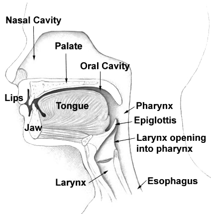
The pharynx (pronounced “FAIR‑inks”) is a hollow, funnel‑shaped organ that forms the posterior wall of the throat. It lies directly behind the mouth (oral cavity) and nasal passages and extends inferiorly to the level of the sixth cervical vertebra, where it bifurcates into the esophagus (for food) and the larynx (for air). Anatomically, the pharynx is divided into three regions: the nasopharynx (above the soft palate), the oropharynx (behind the oral cavity), and the laryngopharynx (or hypopharynx, which continues to the esophagus and larynx). Each segment contains distinct mucosal linings, lymphoid tissue (such as the tonsils), and muscular layers that coordinate swallowing and breathing.During a swallow, a rapid, coordinated sequence of muscle contractions propels the bolus of food posteriorly while the epiglottis, a leaf‑shaped cartilage, folds down to cover the laryngeal inlet, thereby preventing food from entering the airway. Simultaneously, the velopharyngeal sphincter closes off the nasopharynx to stop air from escaping through the nose. The pharynx also participates in speech resonance, immune defense (via the Waldeyer’s ring of lymphoid tissue), and pressure regulation for middle‑ear ventilation through the Eustachian tube.
In vertebrates, the pharynx is a conserved structure, but its morphology varies widely. In fish, it often functions primarily as a conduit for water and prey, while in mammals it has evolved complex muscular and neural control to support both respiration and sophisticated oral processing of food. Even some invertebrates, such as certain mollusks and annelids, possess a pharyngeal chamber that aids in feeding, illustrating the evolutionary versatility of this organ.
History/Background
The term “pharynx” derives from the Greek phárunx, meaning “throat.” Early anatomical descriptions appear in the works of Hippocrates (5th century BC) and Galen (2nd century AD), who recognized the pharynx as a common passage for air and food but lacked detailed knowledge of its muscular coordination. The Renaissance anatomist Andreas Vesalius (1543) provided the first accurate illustrations of the three pharyngeal regions, distinguishing the nasopharynx from the oropharynx. In the 19th century, physiologists such as Claude Bernard and Ivan Pavlov elucidated the reflexive nature of swallowing, identifying the role of the epiglottis and the central pattern generators in the brainstem. The 20th century saw the development of endoscopic techniques, allowing direct visualization of the pharyngeal mucosa and facilitating the diagnosis of disorders like obstructive sleep apnea and pharyngeal cancers. Evolutionary biologists have traced the pharynx’s origins to early chordates, where a simple pharyngeal pouch gave rise to gill slits, later repurposed in terrestrial vertebrates for respiratory and digestive functions.Key Information
- Anatomical divisions: Nasopharynx (air passage, contains the pharyngeal tonsil), Oropharynx (contains palatine and lingual tonsils), Laryngopharynx (connects to esophagus and larynx). - Musculature: Includes the superior, middle, and inferior constrictor muscles, the stylopharyngeus, and the cricopharyngeus (upper esophageal sphincter). - Neural control: Primarily coordinated by the nucleus ambiguus and nucleus tractus solitarius in the medulla, with input from cranial nerves IX (glossopharyngeal), X (vagus), and XII (hypoglossal). - Protective mechanisms: The epiglottis, laryngeal adductor reflex, and the cough reflex protect the lower airway. - Clinical relevance: Dysphagia (difficulty swallowing), aspiration pneumonia, obstructive sleep apnea, and pharyngeal cancers are common concerns; any persistent throat pain, difficulty swallowing, or unexplained weight loss warrants medical evaluation. - Diagnostic tools: Flexible nasopharyngolaryngoscopy, barium swallow studies, and imaging (CT/MRI) assess structural and functional integrity.Significance
The pharynx is essential for survival because it seamlessly integrates two vital life processes—breathing and feeding—into a single anatomical corridor. Its ability to switch rapidly between air and food transport underlies safe swallowing; failure of this system can lead to aspiration, a leading cause of pneumonia, especially in the elderly and neurologically impaired. Moreover, the pharyngeal lymphoid tissue forms a first line of immune defense against inhaled and ingested pathogens, contributing to overall mucosal immunity. In modern medicine, understanding pharyngeal mechanics informs the design of airway management devices (e.g., endotracheal tubes, supraglottic airway masks) and therapeutic interventions for sleep‑disordered breathing. Researchers also study the pharynx’s evolutionary adaptations to gain insight into the transition from aquatic to terrestrial life.When to seek professional care: Persistent sore throat, difficulty swallowing, choking episodes, unexplained hoarseness, or signs of airway obstruction should prompt evaluation by a healthcare professional. Early diagnosis of pharyngeal pathology can prevent complications and improve outcomes.
INFOBOX:
- Name: Pharynx (throat)
- Type: Muscular conduit of the upper aerodigestive tract
- Date: Anatomically described in detail by Andreas Vesalius, 1543 AD
- Location: Posterior to the oral and nasal cavities; extends from the base of the skull to the sixth cervical vertebra
- Known For: Dual transport of air and food, and the epiglottic protection of the airway
TAGS: anatomy, respiratory system, digestive system, swallowing, epiglottis, otolaryngology, evolutionary biology, clinical medicine