Overview
Cartilage is a
specialized connective tissue distinguished by its smooth, glossy appearance and its ability to withstand compressive forces while remaining flexible. Unlike bone, cartilage lacks a mineralized matrix and blood vessels; instead, it is nourished by diffusion from the surrounding
perichondrium, a tough fibrous membrane that envelops most cartilage types. The tissue is composed of a dense extracellular matrix rich in
glycosaminoglycans (GAGs),
proteoglycans,
collagen fibers (predominantly type II), and, in certain varieties,
elastin. This matrix provides cartilage with its characteristic combination of stiffness and elasticity—stiffer than muscle or tendon but far more pliable than bone.
In tetrapods, cartilage performs several critical roles. Articular cartilage coats the ends of long bones within synovial joints, reducing friction and absorbing shock during movement. It also forms structural components of the rib cage, larynx, bronchial tubes, and intervertebral discs, where it contributes to spinal flexibility and load distribution. In chondrichthyans (sharks, rays) and cyclostomes (lampreys, hagfish), cartilage replaces bone as the dominant skeletal element, providing a lightweight yet sturdy framework for the entire organism. Because cartilage is avascular, it heals slowly; injuries to articular surfaces often require medical evaluation, and persistent pain or swelling should prompt a visit to a healthcare professional.
History/Background
The scientific study of cartilage dates back to the early anatomical investigations of the Renaissance. Leonardo da Vinci’s sketches (c. 1490) noted the “white, glassy” nature of joint surfaces, while Andreas Vesalius (1543) described the perichondrium in his seminal work
De humani corporis fabrica. In the 19th century,
Rudolf Virchow coined the term “chondrification” to describe the process by which mesenchymal cells differentiate into cartilage‑producing chondroblasts. The discovery of
collagen type II in the 1950s and the elucidation of the
glycosaminoglycan–proteoglycan complex in the 1960s clarified the molecular basis of cartilage’s mechanical properties. More recently, advances in tissue engineering (2000s‑2020s) have focused on cultivating
autologous chondrocyte implants and
3‑D‑printed scaffolds to repair damaged articular cartilage, reflecting a shift from descriptive anatomy to regenerative therapeutics.
Key Information
-
Types of cartilage: hyaline (most common, found in joints and respiratory tract), fibrocartilage (dense collagen, present in intervertebral discs and menisci), and elastic cartilage (high elastin content, forms the ear pinna and epiglottis).
-
Matrix composition: ~70‑80 % water, 20‑30 % solid matrix; GAGs such as chondroitin sulfate and keratan sulfate attract water, creating a hydrogel that resists compression.
-
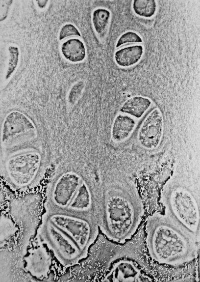
Cellular components: chondrocytes reside in lacunae, maintaining the matrix; they originate from mesenchymal stem cells and exhibit limited proliferative capacity in mature tissue.
-
Growth and repair: cartilage grows faster than bone during childhood, primarily through
endochondral ossification where a cartilage template is later replaced by bone. In adults, repair relies on diffusion and limited chondrocyte activity; thus, injuries often progress to osteoarthritis if untreated.
-
Clinical relevance: degeneration of articular cartilage is the hallmark of osteoarthritis; traumatic lesions may require arthroscopic debridement, microfracture, or cell‑based therapies.
-
Comparative anatomy: in sharks, the skeleton consists of
calcified cartilage reinforced with mineral deposits, offering a model for bio‑inspired materials.
Significance
Cartilage’s unique biomechanical profile makes it indispensable for locomotion, respiration, and structural integrity across vertebrate species. Its ability to absorb shock protects bone surfaces from wear, while its flexibility enables complex movements such as bending of the spine and articulation of the jaw. In medicine, understanding cartilage biology underpins the treatment of joint disorders, sports injuries, and age‑related degeneration. The ongoing development of
regenerative cartilage therapies promises to reduce the burden of osteoarthritis, a leading cause of disability worldwide. Moreover, the study of cartilaginous skeletons in chondrichthyans informs evolutionary biology and inspires engineering of lightweight, high‑strength composites. For anyone experiencing persistent joint pain, swelling, or reduced range of motion, seeking professional evaluation is essential, as early intervention can preserve cartilage health and prevent long‑term disability.