



Dr. Sage Newton
Science Editor
241Articles
2.8KTotal views
 Science
Cell Membrane
** The cell membrane is a dynamic, semipermeable lipid bilayer that encloses the cell, regulates molecular traffic, and mediates communication with the external environment.
**CONTENT:**
## Overview
The **cell membrane**—also called the plasma membrane—is a thin, flexible barrier that defines the boundary of every living cell. Composed primarily of a **lipid bilayer**, it consists of two opposing sheets of phospholipids whose hydrophilic heads face outward toward the aqueous extracellular and intracellular fluids, while their hydrophobic tails tuck inward, creating a water‑impermeable core. Interspersed among these phospholipids are **glycolipids**, which bear carbohydrate chains that participate in cell‑cell recognition, and, in most eukaryotes and many archaea, **sterols** such as cholesterol (≈30 % of total lipids in human erythrocytes) that modulate membrane fluidity across temperature ranges from 0 °C to 40 °C.
Embedded within this fluid matrix are **membrane proteins** that fall into two major classes. **Integral (or transmembrane) proteins** span the bilayer one or more times, forming channels, carriers, and receptors that control the selective passage of ions, nutrients, and signaling molecules. **Peripheral proteins** loosely associate with the inner or outer leaflets, often acting as enzymes, scaffolds, or anchors for the cytoskeleton. Together, these components create a highly organized, yet adaptable, platform that enables cells to maintain homeostasis, sense their surroundings, and interact with other cells.
The membrane’s **semipermeable** nature means that while small, non‑polar molecules (e.g., O₂, CO₂) diffuse freely, larger or charged species require specialized transport mechanisms. This selective permeability is quantified by the **permeability coefficient (P)**, which for water across a typical mammalian plasma membrane is about 10⁻³ cm s⁻¹ at 37 °C. The dynamic rearrangement of lipids and proteins—often described by the **fluid‑ mosaic model**—allows the membrane to undergo rapid remodeling during processes such as endocytosis, exocytosis, and cell division.
## History/Background
The concept of a distinct cellular boundary dates back to **Robert Hooke’s** 1665 observations of “cells” in cork, but the biochemical nature of the membrane remained elusive for centuries. In **1925**, **Gorter and Grendel** extracted lipids from red blood cells and, using the **Langmuir trough**, measured a surface area roughly twice that of the cells, leading to the first proposal of a **lipid bilayer**. The seminal **Davson‑Danielli model (1935)** suggested a protein “sandwich” surrounding the lipid layer, a view later overturned by **S.J. Singer and Garth L. Nicolson’s fluid‑mosaic model (1972)**, which integrated the roles of proteins and lipids into a unified, dynamic structure. Advances in electron microscopy (1970s) and later **cryo‑electron tomography** (2000s) provided visual confirmation of the bilayer’s thickness (~5 nm) and protein distribution. The discovery of **cholesterol’s** stabilizing effect in the 1970s and the identification of **glycolipid-mediated cell‑recognition** pathways in the 1980s further refined our understanding of membrane complexity.
## Key Information
- **Composition:** ~50 % phospholipids, 30 % cholesterol (in animal cells), 10 % glycolipids, 10 % proteins (by mass).
- **Thickness:** ~5 nm (≈ 50 Å).
- **Fluidity:** Modulated by fatty‑acid saturation; unsaturated chains increase fluidity, while sterols decrease it at high temperatures and prevent solidification at low temperatures.
- **Transport mechanisms:**
* **Passive diffusion** (small, non‑polar molecules).
* **Facilitated diffusion** via channel proteins (e.g., aquaporins, with water permeability up to 10⁹ water molecules s⁻¹ per channel).
* **Active transport** using ATP‑driven pumps (e.g., Na⁺/K⁺‑ATPase moves 3 Na⁺ out and 2 K⁺ in per ATP hydrolyzed).
* **Endocytosis/exocytosis** for macromolecule uptake and secretion.
- **Signal transduction:** Receptor tyrosine kinases (RTKs) and G‑protein‑coupled receptors (GPCRs) reside in the membrane, converting extracellular cues into intracellular responses within milliseconds.
- **Membrane potential:** Typically –70 mV in resting mammalian neurons, generated by ion gradients maintained by pumps and leak channels.
## Significance
The cell membrane is central to **cellular life**; without it, the delicate balance of ions, metabolites, and signaling molecules would collapse, leading to loss of function and viability. Its **selective permeability** underpins nerve impulse propagation, muscle contraction, and hormone signaling, while its **receptor repertoire** enables organisms to respond to nutrients, toxins, and pathogens. In medicine, membrane proteins constitute **≈ 60 % of all drug targets**, exemplified by antihypertensives (β‑blockers) and antibiotics (β‑lactams) that bind to specific membrane components. Moreover, engineered **lipid nanoparticles**—leveraging the natural fusogenic properties of membranes—have become the delivery vehicles for mRNA vaccines (e.g., COVID‑19 vaccines approved in 2020). Understanding membrane dynamics also informs synthetic biology, where artificial vesicles mimic cellular compartments for bioreactors and drug screening.
**INFOBOX:**
- Name: Plasma (Cell) Membrane
- Type: Biological membrane (semipermeable barrier)
- Date: First described as a lipid bilayer in 1925 (Gorter & Grendel)
- Location: Encloses every living cell, from bacteria to human neurons
- Known For: Regulating transport, signaling, and structural integrity of cells
**TAGS:** cell biology, membrane biophysics, lipid bilayer, membrane proteins, fluid mosaic model, signal transduction, drug targets, biotechnology
Science
Cell Membrane
** The cell membrane is a dynamic, semipermeable lipid bilayer that encloses the cell, regulates molecular traffic, and mediates communication with the external environment.
**CONTENT:**
## Overview
The **cell membrane**—also called the plasma membrane—is a thin, flexible barrier that defines the boundary of every living cell. Composed primarily of a **lipid bilayer**, it consists of two opposing sheets of phospholipids whose hydrophilic heads face outward toward the aqueous extracellular and intracellular fluids, while their hydrophobic tails tuck inward, creating a water‑impermeable core. Interspersed among these phospholipids are **glycolipids**, which bear carbohydrate chains that participate in cell‑cell recognition, and, in most eukaryotes and many archaea, **sterols** such as cholesterol (≈30 % of total lipids in human erythrocytes) that modulate membrane fluidity across temperature ranges from 0 °C to 40 °C.
Embedded within this fluid matrix are **membrane proteins** that fall into two major classes. **Integral (or transmembrane) proteins** span the bilayer one or more times, forming channels, carriers, and receptors that control the selective passage of ions, nutrients, and signaling molecules. **Peripheral proteins** loosely associate with the inner or outer leaflets, often acting as enzymes, scaffolds, or anchors for the cytoskeleton. Together, these components create a highly organized, yet adaptable, platform that enables cells to maintain homeostasis, sense their surroundings, and interact with other cells.
The membrane’s **semipermeable** nature means that while small, non‑polar molecules (e.g., O₂, CO₂) diffuse freely, larger or charged species require specialized transport mechanisms. This selective permeability is quantified by the **permeability coefficient (P)**, which for water across a typical mammalian plasma membrane is about 10⁻³ cm s⁻¹ at 37 °C. The dynamic rearrangement of lipids and proteins—often described by the **fluid‑ mosaic model**—allows the membrane to undergo rapid remodeling during processes such as endocytosis, exocytosis, and cell division.
## History/Background
The concept of a distinct cellular boundary dates back to **Robert Hooke’s** 1665 observations of “cells” in cork, but the biochemical nature of the membrane remained elusive for centuries. In **1925**, **Gorter and Grendel** extracted lipids from red blood cells and, using the **Langmuir trough**, measured a surface area roughly twice that of the cells, leading to the first proposal of a **lipid bilayer**. The seminal **Davson‑Danielli model (1935)** suggested a protein “sandwich” surrounding the lipid layer, a view later overturned by **S.J. Singer and Garth L. Nicolson’s fluid‑mosaic model (1972)**, which integrated the roles of proteins and lipids into a unified, dynamic structure. Advances in electron microscopy (1970s) and later **cryo‑electron tomography** (2000s) provided visual confirmation of the bilayer’s thickness (~5 nm) and protein distribution. The discovery of **cholesterol’s** stabilizing effect in the 1970s and the identification of **glycolipid-mediated cell‑recognition** pathways in the 1980s further refined our understanding of membrane complexity.
## Key Information
- **Composition:** ~50 % phospholipids, 30 % cholesterol (in animal cells), 10 % glycolipids, 10 % proteins (by mass).
- **Thickness:** ~5 nm (≈ 50 Å).
- **Fluidity:** Modulated by fatty‑acid saturation; unsaturated chains increase fluidity, while sterols decrease it at high temperatures and prevent solidification at low temperatures.
- **Transport mechanisms:**
* **Passive diffusion** (small, non‑polar molecules).
* **Facilitated diffusion** via channel proteins (e.g., aquaporins, with water permeability up to 10⁹ water molecules s⁻¹ per channel).
* **Active transport** using ATP‑driven pumps (e.g., Na⁺/K⁺‑ATPase moves 3 Na⁺ out and 2 K⁺ in per ATP hydrolyzed).
* **Endocytosis/exocytosis** for macromolecule uptake and secretion.
- **Signal transduction:** Receptor tyrosine kinases (RTKs) and G‑protein‑coupled receptors (GPCRs) reside in the membrane, converting extracellular cues into intracellular responses within milliseconds.
- **Membrane potential:** Typically –70 mV in resting mammalian neurons, generated by ion gradients maintained by pumps and leak channels.
## Significance
The cell membrane is central to **cellular life**; without it, the delicate balance of ions, metabolites, and signaling molecules would collapse, leading to loss of function and viability. Its **selective permeability** underpins nerve impulse propagation, muscle contraction, and hormone signaling, while its **receptor repertoire** enables organisms to respond to nutrients, toxins, and pathogens. In medicine, membrane proteins constitute **≈ 60 % of all drug targets**, exemplified by antihypertensives (β‑blockers) and antibiotics (β‑lactams) that bind to specific membrane components. Moreover, engineered **lipid nanoparticles**—leveraging the natural fusogenic properties of membranes—have become the delivery vehicles for mRNA vaccines (e.g., COVID‑19 vaccines approved in 2020). Understanding membrane dynamics also informs synthetic biology, where artificial vesicles mimic cellular compartments for bioreactors and drug screening.
**INFOBOX:**
- Name: Plasma (Cell) Membrane
- Type: Biological membrane (semipermeable barrier)
- Date: First described as a lipid bilayer in 1925 (Gorter & Grendel)
- Location: Encloses every living cell, from bacteria to human neurons
- Known For: Regulating transport, signaling, and structural integrity of cells
**TAGS:** cell biology, membrane biophysics, lipid bilayer, membrane proteins, fluid mosaic model, signal transduction, drug targets, biotechnology
 Science
Microbiology
** Microbiology is the scientific discipline devoted to the study of microorganisms—organisms too small to be seen unaided—including bacteria, archaea, viruses, fungi, protozoa, and microscopic algae.
**CONTENT:**
## Overview
Microbiology explores life at the smallest scales, revealing how single‑celled and acellular entities drive ecosystems, human health, and industrial processes. These microorganisms can be **unicellular**, **multicellular**, or even **acellular** (as in viruses), and they inhabit every conceivable niche—from the icy depths of Antarctic lakes to the scorching vents of hydrothermal chimneys. Modern microbiologists employ a blend of classical culture techniques, molecular genetics, and high‑throughput sequencing to decipher microbial metabolism, genetics, and interactions.
The field is inherently interdisciplinary, intersecting **biochemistry**, **ecology**, **medicine**, and **engineering**. Sub‑disciplines such as **virology** (study of viruses), **bacteriology** (bacteria), **protistology** (protozoa and related eukaryotes), **mycology** (fungi), **immunology** (host‑microbe interactions), and **parasitology** (parasitic organisms) each focus on distinct groups but share common tools and concepts. By understanding microbes, scientists harness them for **bioremediation**, **biotechnology**, **food production**, and **pharmaceutical development**, while also devising strategies to combat infectious diseases.
## History/Background
The roots of microbiology trace back to the invention of the microscope in the late 16th century. In 1676, **Antonie van Leeuwenhoek** reported “animalcules” observed in pond water, marking the first documented glimpse of microorganisms. The 19th century ushered in a scientific framework: **Louis Pasteur** (1822‑1895) demonstrated that microbes cause fermentation and disease, establishing the **germ theory of disease** in 1861, while **Robert Koch** (1843‑1910) formulated Koch’s postulates (1884) to link specific microbes to specific illnesses.
The early 20th century saw the rise of **bacteriology** as a clinical discipline, with the discovery of antibiotics—penicillin by **Alexander Fleming** in 1928—revolutionizing treatment. **Virology** emerged after the identification of the tobacco mosaic virus in 1892 and the subsequent isolation of the first animal virus (foot‑and‑mouth disease) in 1898. The mid‑20th century brought molecular tools: the **DNA double‑helix** model (1953) and the development of **polymerase chain reaction (PCR)** in 1983, enabling rapid microbial identification. In the 21st century, **metagenomics** and **next‑generation sequencing** have uncovered that over 99 % of microbial species remain uncultured, expanding the known tree of life dramatically.
## Key Information
- **Domains of life:** Microbiology studies organisms across the three domains—**Bacteria**, **Archaea**, and **Eukarya** (including fungi and protists).
- **Population size:** An average gram of soil contains **10⁹–10¹⁰** bacterial cells; the human gut hosts roughly **10¹⁴** microbes, outnumbering human cells 10‑to‑1.
- **Techniques:** Classic plate culturing, **Gram staining**, **electron microscopy**, **16S rRNA gene sequencing**, and **CRISPR‑Cas genome editing** are core methods.
- **Achievements:** Development of vaccines (e.g., polio, 1955), discovery of **DNA replication enzymes** (DNA polymerase I, 1958), and engineering of **synthetic biology chassis** like *E. coli* for bio‑fuel production.
- **Applications:** Wastewater treatment via **activated sludge**, production of antibiotics (e.g., streptomycin, 1943), fermentation of foods (yogurt, cheese), and bioprocessing of enzymes (amylase, cellulase).
## Significance
Microbiology underpins modern medicine, agriculture, and industry. Understanding pathogenic microbes informs **public health policies**, as seen during the COVID‑19 pandemic (SARS‑CoV‑2 identified in January 2020). In agriculture, **soil microbiomes** enhance nutrient cycling, reducing reliance on synthetic fertilizers. Industrially, microbes are bio‑factories: engineered yeast produces the antimalarial drug artemisinin at scale, and cyanobacteria are being trialed for carbon capture.
Beyond practical uses, microbiology reshapes our philosophical view of life. The discovery that microbes exchange genes horizontally challenges the classic tree‑like model of evolution, suggesting a **network of genetic exchange**. Moreover, the recognition that microbial processes regulate planetary biogeochemical cycles—such as the **carbon** and **nitrogen cycles**—highlights microbes as essential architects of Earth’s climate system. As humanity confronts climate change, antibiotic resistance, and emerging pathogens, microbiology offers tools and insights vital for sustainable solutions.
**INFOBOX:**
- Name: Microbiology
- Type: Scientific discipline (biological science)
- Date: Established as a formal field in the late 19th century (germ theory, 1860s)
- Location: Global (research institutions, universities, industry labs)
- Known For: Study of microorganisms and their applications in health, industry, and ecology
**TAGS:** microbiology, bacteria, viruses, immunology, biotechnology, infectious disease, ecology, molecular biology
Science
Microbiology
** Microbiology is the scientific discipline devoted to the study of microorganisms—organisms too small to be seen unaided—including bacteria, archaea, viruses, fungi, protozoa, and microscopic algae.
**CONTENT:**
## Overview
Microbiology explores life at the smallest scales, revealing how single‑celled and acellular entities drive ecosystems, human health, and industrial processes. These microorganisms can be **unicellular**, **multicellular**, or even **acellular** (as in viruses), and they inhabit every conceivable niche—from the icy depths of Antarctic lakes to the scorching vents of hydrothermal chimneys. Modern microbiologists employ a blend of classical culture techniques, molecular genetics, and high‑throughput sequencing to decipher microbial metabolism, genetics, and interactions.
The field is inherently interdisciplinary, intersecting **biochemistry**, **ecology**, **medicine**, and **engineering**. Sub‑disciplines such as **virology** (study of viruses), **bacteriology** (bacteria), **protistology** (protozoa and related eukaryotes), **mycology** (fungi), **immunology** (host‑microbe interactions), and **parasitology** (parasitic organisms) each focus on distinct groups but share common tools and concepts. By understanding microbes, scientists harness them for **bioremediation**, **biotechnology**, **food production**, and **pharmaceutical development**, while also devising strategies to combat infectious diseases.
## History/Background
The roots of microbiology trace back to the invention of the microscope in the late 16th century. In 1676, **Antonie van Leeuwenhoek** reported “animalcules” observed in pond water, marking the first documented glimpse of microorganisms. The 19th century ushered in a scientific framework: **Louis Pasteur** (1822‑1895) demonstrated that microbes cause fermentation and disease, establishing the **germ theory of disease** in 1861, while **Robert Koch** (1843‑1910) formulated Koch’s postulates (1884) to link specific microbes to specific illnesses.
The early 20th century saw the rise of **bacteriology** as a clinical discipline, with the discovery of antibiotics—penicillin by **Alexander Fleming** in 1928—revolutionizing treatment. **Virology** emerged after the identification of the tobacco mosaic virus in 1892 and the subsequent isolation of the first animal virus (foot‑and‑mouth disease) in 1898. The mid‑20th century brought molecular tools: the **DNA double‑helix** model (1953) and the development of **polymerase chain reaction (PCR)** in 1983, enabling rapid microbial identification. In the 21st century, **metagenomics** and **next‑generation sequencing** have uncovered that over 99 % of microbial species remain uncultured, expanding the known tree of life dramatically.
## Key Information
- **Domains of life:** Microbiology studies organisms across the three domains—**Bacteria**, **Archaea**, and **Eukarya** (including fungi and protists).
- **Population size:** An average gram of soil contains **10⁹–10¹⁰** bacterial cells; the human gut hosts roughly **10¹⁴** microbes, outnumbering human cells 10‑to‑1.
- **Techniques:** Classic plate culturing, **Gram staining**, **electron microscopy**, **16S rRNA gene sequencing**, and **CRISPR‑Cas genome editing** are core methods.
- **Achievements:** Development of vaccines (e.g., polio, 1955), discovery of **DNA replication enzymes** (DNA polymerase I, 1958), and engineering of **synthetic biology chassis** like *E. coli* for bio‑fuel production.
- **Applications:** Wastewater treatment via **activated sludge**, production of antibiotics (e.g., streptomycin, 1943), fermentation of foods (yogurt, cheese), and bioprocessing of enzymes (amylase, cellulase).
## Significance
Microbiology underpins modern medicine, agriculture, and industry. Understanding pathogenic microbes informs **public health policies**, as seen during the COVID‑19 pandemic (SARS‑CoV‑2 identified in January 2020). In agriculture, **soil microbiomes** enhance nutrient cycling, reducing reliance on synthetic fertilizers. Industrially, microbes are bio‑factories: engineered yeast produces the antimalarial drug artemisinin at scale, and cyanobacteria are being trialed for carbon capture.
Beyond practical uses, microbiology reshapes our philosophical view of life. The discovery that microbes exchange genes horizontally challenges the classic tree‑like model of evolution, suggesting a **network of genetic exchange**. Moreover, the recognition that microbial processes regulate planetary biogeochemical cycles—such as the **carbon** and **nitrogen cycles**—highlights microbes as essential architects of Earth’s climate system. As humanity confronts climate change, antibiotic resistance, and emerging pathogens, microbiology offers tools and insights vital for sustainable solutions.
**INFOBOX:**
- Name: Microbiology
- Type: Scientific discipline (biological science)
- Date: Established as a formal field in the late 19th century (germ theory, 1860s)
- Location: Global (research institutions, universities, industry labs)
- Known For: Study of microorganisms and their applications in health, industry, and ecology
**TAGS:** microbiology, bacteria, viruses, immunology, biotechnology, infectious disease, ecology, molecular biology
 Science
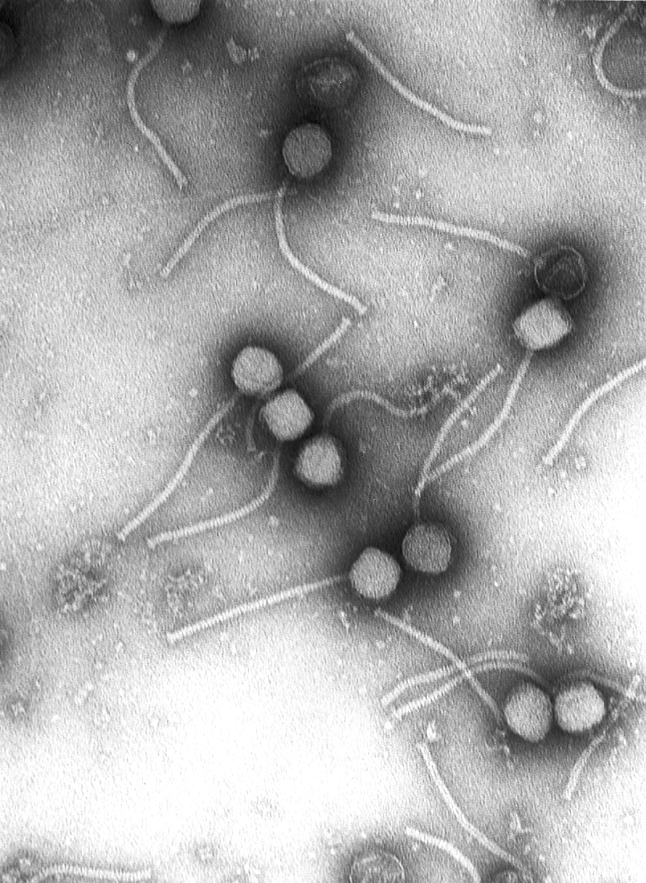
Virology
** Virology is the scientific discipline that investigates viruses—their structure, genetics, life cycles, interactions with hosts, and applications in medicine and biotechnology.
**CONTENT:**
## Overview
Virology sits at the intersection of **microbiology**, **molecular biology**, and **immunology**, focusing on entities that straddle the line between living and non‑living. Viruses are obligate intracellular parasites; they consist of nucleic acid (either **DNA** or **RNA**) encased in a protein **capsid**, and in many cases a lipid **envelope** derived from host membranes. Their sizes range from ~20 nm for the smallest **parvoviruses** to >300 nm for giant **Mimiviruses**, and a single virion can contain from a few thousand to several hundred thousand base pairs of genetic material.
The field examines how viruses **detect**, **enter**, and **replicate** within host cells, exploiting cellular machinery to produce progeny virions—often at rates exceeding 10^9 particles per infected cell within hours. Virologists also study viral **evolution**, which proceeds at rates up to 10^−3 substitutions per site per year for RNA viruses, far faster than most cellular organisms. This rapid evolution underlies the emergence of new pathogens, vaccine escape mutants, and antiviral resistance.
Beyond disease, viruses are powerful tools. Engineered **viral vectors** deliver therapeutic genes in gene therapy, while bacteriophages are being revived as precision antibiotics. The discipline therefore bridges fundamental science with translational applications, from pandemic preparedness to synthetic biology.
## History/Background
The roots of virology trace back to the late 19th century. In 1892, Dmitri **Ivanovsky** demonstrated that the agent causing tobacco mosaic disease could pass through porcelain filters that retained bacteria, hinting at a “filterable” pathogen. Martin **Beijerinck** coined the term **virus** in 1898, describing it as a “contagium vivum fluidum.” The first animal virus, **foot‑and‑mouth disease virus**, was isolated in 1898, and the first human virus, **yellow fever virus**, was identified in 1901.
The 20th century saw rapid methodological advances. The invention of the **electron microscope** in the 1930s allowed direct visualization of virions, confirming their particulate nature. In 1952, **Alfred Hershey** and **Martha Chase** used radiolabeled phage to prove that DNA, not protein, carries genetic information—a cornerstone of molecular genetics. The discovery of the **reverse transcriptase** enzyme in 1970 (Howard Temin, David Baltimore) revealed that RNA viruses could integrate into host genomes, reshaping our understanding of genetic flow.
The **Molecular Era** began in the 1980s with recombinant DNA technology, enabling the cloning of viral genomes and the creation of attenuated vaccines (e.g., measles, mumps, rubella). The 2000s brought high‑throughput **next‑generation sequencing**, allowing real‑time tracking of viral outbreaks, exemplified by the rapid sequencing of the **SARS‑CoV‑2** genome in January 2020—completed within weeks of the first reported cases.
## Key Information
- **Classification:** Viruses are grouped by nucleic acid type (DNA vs. RNA), strandedness (single vs. double), sense (positive vs. negative), envelope presence, and replication strategy (the **Baltimore classification**, six groups).
- **Detection & Isolation:** Techniques include **PCR**, **RT‑PCR**, **ELISA**, **viral culture** in cell lines (e.g., Vero, MDCK), and **plaque assays** to quantify infectious units (plaque‑forming units, PFU).
- **Life Cycles:** Canonical cycles include **attachment**, **penetration**, **uncoating**, **replication**, **assembly**, and **release** (via lysis or budding). Some viruses, like **herpesviruses**, establish latency, persisting in host cells for life.
- **Therapeutics:** Antiviral drugs target specific stages—**acyclovir** inhibits viral DNA polymerase; **oseltamivir** blocks neuraminidase in influenza. **Vaccines** (live‑attenuated, inactivated, subunit, mRNA) have eradicated smallpox (1980) and dramatically reduced polio incidence.
- **Research Tools:** **Bacteriophages** serve as model systems for genetics; **adenoviral vectors** deliver CRISPR components; **virus‑like particles (VLPs)** provide safe immunogens.
## Significance
Virology is pivotal to global health. Emerging infections—HIV (identified 1983), Ebola (1976), Zika (2015), and COVID‑19 (2020)—have underscored the need for rapid viral detection, surveillance, and vaccine development. Understanding viral mechanisms informs **immune system** research, revealing how innate sensors (e.g., **RIG‑I**, **cGAS**) detect foreign nucleic acids and trigger interferon responses.
Economically, the field drives billions in pharmaceutical revenue; the mRNA vaccine platform, honed through decades of virology, delivered >10 billion doses of COVID‑19 vaccines within two years, showcasing the translational power of viral research. Moreover, phage therapy offers a sustainable alternative to antibiotics amid rising antimicrobial resistance.
In ecological terms, viruses regulate microbial populations, influence nutrient cycles, and shape evolutionary trajectories across all domains of life. Their ubiquity—estimated 10^31 virions on Earth—makes them the most abundant biological entities, a fact that continues to inspire new scientific frontiers, from **viral dark matter** metagenomics to synthetic virology.
**INFOBOX:**
- Name: Virology
- Type: Scientific discipline (subfield of microbiology)
- Date: Established as a distinct field in the late 19th century (circa 1892)
- Location: Global (research institutions, universities, public health labs)
- Known For: Study of virus structure, replication, pathogenesis, and application in medicine and biotechnology
**TAGS:** virology, viruses, microbiology, immunology, molecular biology, infectious disease, vaccine development, gene therapy
Science
Virology
** Virology is the scientific discipline that investigates viruses—their structure, genetics, life cycles, interactions with hosts, and applications in medicine and biotechnology.
**CONTENT:**
## Overview
Virology sits at the intersection of **microbiology**, **molecular biology**, and **immunology**, focusing on entities that straddle the line between living and non‑living. Viruses are obligate intracellular parasites; they consist of nucleic acid (either **DNA** or **RNA**) encased in a protein **capsid**, and in many cases a lipid **envelope** derived from host membranes. Their sizes range from ~20 nm for the smallest **parvoviruses** to >300 nm for giant **Mimiviruses**, and a single virion can contain from a few thousand to several hundred thousand base pairs of genetic material.
The field examines how viruses **detect**, **enter**, and **replicate** within host cells, exploiting cellular machinery to produce progeny virions—often at rates exceeding 10^9 particles per infected cell within hours. Virologists also study viral **evolution**, which proceeds at rates up to 10^−3 substitutions per site per year for RNA viruses, far faster than most cellular organisms. This rapid evolution underlies the emergence of new pathogens, vaccine escape mutants, and antiviral resistance.
Beyond disease, viruses are powerful tools. Engineered **viral vectors** deliver therapeutic genes in gene therapy, while bacteriophages are being revived as precision antibiotics. The discipline therefore bridges fundamental science with translational applications, from pandemic preparedness to synthetic biology.
## History/Background
The roots of virology trace back to the late 19th century. In 1892, Dmitri **Ivanovsky** demonstrated that the agent causing tobacco mosaic disease could pass through porcelain filters that retained bacteria, hinting at a “filterable” pathogen. Martin **Beijerinck** coined the term **virus** in 1898, describing it as a “contagium vivum fluidum.” The first animal virus, **foot‑and‑mouth disease virus**, was isolated in 1898, and the first human virus, **yellow fever virus**, was identified in 1901.
The 20th century saw rapid methodological advances. The invention of the **electron microscope** in the 1930s allowed direct visualization of virions, confirming their particulate nature. In 1952, **Alfred Hershey** and **Martha Chase** used radiolabeled phage to prove that DNA, not protein, carries genetic information—a cornerstone of molecular genetics. The discovery of the **reverse transcriptase** enzyme in 1970 (Howard Temin, David Baltimore) revealed that RNA viruses could integrate into host genomes, reshaping our understanding of genetic flow.
The **Molecular Era** began in the 1980s with recombinant DNA technology, enabling the cloning of viral genomes and the creation of attenuated vaccines (e.g., measles, mumps, rubella). The 2000s brought high‑throughput **next‑generation sequencing**, allowing real‑time tracking of viral outbreaks, exemplified by the rapid sequencing of the **SARS‑CoV‑2** genome in January 2020—completed within weeks of the first reported cases.
## Key Information
- **Classification:** Viruses are grouped by nucleic acid type (DNA vs. RNA), strandedness (single vs. double), sense (positive vs. negative), envelope presence, and replication strategy (the **Baltimore classification**, six groups).
- **Detection & Isolation:** Techniques include **PCR**, **RT‑PCR**, **ELISA**, **viral culture** in cell lines (e.g., Vero, MDCK), and **plaque assays** to quantify infectious units (plaque‑forming units, PFU).
- **Life Cycles:** Canonical cycles include **attachment**, **penetration**, **uncoating**, **replication**, **assembly**, and **release** (via lysis or budding). Some viruses, like **herpesviruses**, establish latency, persisting in host cells for life.
- **Therapeutics:** Antiviral drugs target specific stages—**acyclovir** inhibits viral DNA polymerase; **oseltamivir** blocks neuraminidase in influenza. **Vaccines** (live‑attenuated, inactivated, subunit, mRNA) have eradicated smallpox (1980) and dramatically reduced polio incidence.
- **Research Tools:** **Bacteriophages** serve as model systems for genetics; **adenoviral vectors** deliver CRISPR components; **virus‑like particles (VLPs)** provide safe immunogens.
## Significance
Virology is pivotal to global health. Emerging infections—HIV (identified 1983), Ebola (1976), Zika (2015), and COVID‑19 (2020)—have underscored the need for rapid viral detection, surveillance, and vaccine development. Understanding viral mechanisms informs **immune system** research, revealing how innate sensors (e.g., **RIG‑I**, **cGAS**) detect foreign nucleic acids and trigger interferon responses.
Economically, the field drives billions in pharmaceutical revenue; the mRNA vaccine platform, honed through decades of virology, delivered >10 billion doses of COVID‑19 vaccines within two years, showcasing the translational power of viral research. Moreover, phage therapy offers a sustainable alternative to antibiotics amid rising antimicrobial resistance.
In ecological terms, viruses regulate microbial populations, influence nutrient cycles, and shape evolutionary trajectories across all domains of life. Their ubiquity—estimated 10^31 virions on Earth—makes them the most abundant biological entities, a fact that continues to inspire new scientific frontiers, from **viral dark matter** metagenomics to synthetic virology.
**INFOBOX:**
- Name: Virology
- Type: Scientific discipline (subfield of microbiology)
- Date: Established as a distinct field in the late 19th century (circa 1892)
- Location: Global (research institutions, universities, public health labs)
- Known For: Study of virus structure, replication, pathogenesis, and application in medicine and biotechnology
**TAGS:** virology, viruses, microbiology, immunology, molecular biology, infectious disease, vaccine development, gene therapy
 People
Roger Penrose
Sir Roger Penrose is a British mathematician and physicist whose revolutionary work on black holes, spacetime singularities, and geometric tilings has fundamentally reshaped our understanding of the universe and inspired fields from cosmology to materials science.
People
Roger Penrose
Sir Roger Penrose is a British mathematician and physicist whose revolutionary work on black holes, spacetime singularities, and geometric tilings has fundamentally reshaped our understanding of the universe and inspired fields from cosmology to materials science.
 Science
Evolutionary Biology
** Evolutionary biology is the scientific discipline that investigates how genetic variation arises and spreads through natural selection, mutation, genetic drift, and gene flow, shaping the diversity of life on Earth.
**CONTENT:**
## Overview
Evolutionary biology sits at the heart of the life sciences, providing a unifying framework for understanding the origin, adaptation, and extinction of organisms. By integrating genetics, ecology, paleontology, and developmental biology, the field explains how **heritable traits** change in frequency within populations over generations. Central to this discipline are four mechanisms of evolution: **natural selection**, the differential survival and reproduction of individuals; **mutation**, the ultimate source of new genetic variation; **genetic drift**, random fluctuations in allele frequencies especially in small populations; and **gene flow**, the transfer of genes among populations through migration.
The modern perspective treats evolution as a dynamic, measurable process. Researchers employ mathematical models, genome sequencing, and experimental evolution (e.g., long‑term *E. coli* cultures) to quantify rates of change, often expressed in **selection coefficients** (s) ranging from 0.001 to 0.1 per generation. These tools allow scientists to predict evolutionary trajectories, assess the impact of climate change on species, and even guide medical strategies such as antibiotic stewardship.
## History/Background
The intellectual roots of evolutionary biology trace back to the mid‑19th century. In 1858, **Charles Darwin** and **Alfred Russel Wallace** independently presented the concept of natural selection, drawing on biogeographic patterns such as the distinct finches of the Galápagos Islands. Their joint paper was published in *The Journal of the Proceedings of the Linnean Society* (1858), and Darwin’s seminal work, *On the Origin of Species* (1859), laid the groundwork for a mechanistic view of evolution.
Parallel to these ideas, **Gregor Mendel** (1865) uncovered the laws of inheritance through pea‑plant experiments, establishing the particulate nature of genetic transmission. For decades, Darwin’s and Mendel’s insights seemed incompatible. The reconciliation arrived in the early 20th century with **Ronald A. Fisher**, **J.B.S. Haldane**, and **Sewall Wright**, who forged the **Modern Synthesis** (1930s–1940s). Fisher’s 1930 book *The Genetical Theory of Natural Selection* mathematically merged selection with Mendelian genetics, cementing a unified theory that still underpins evolutionary biology today.
## Key Information
- **Four mechanisms of evolution**: natural selection, mutation, genetic drift, gene flow.
- **Natural selection**: first articulated by Darwin and Wallace (1858–1859); quantified by fitness differentials (e.g., a 5 % advantage per generation yields a selection coefficient s = 0.05).
- **Mutation rate**: in humans ≈ 1 × 10⁻⁸ mutations per nucleotide per generation; in microbes up to 10⁻⁶.
- **Genetic drift**: strongest in populations < 1,000 individuals; the probability of fixation of a neutral allele equals its initial frequency.
- **Gene flow**: measured by migration rate (m); even m ≈ 0.01 can homogenize allele frequencies across subpopulations.
- **Key figures**: Darwin (1809–1882), Wallace (1823–1913), Mendel (1822–1884), Fisher (1890–1962).
- **Major achievements**: elucidation of adaptive radiations, discovery of DNA’s double helix (1953) linking genotype to phenotype, development of phylogenomic methods that reconstruct the tree of life with > 100,000 species sampled.
- **Tools**: population‑genetic models (e.g., Wright–Fisher), comparative genomics, experimental evolution, fossil calibration (e.g., Cambrian explosion ~541 Ma).
## Significance
Evolutionary biology provides the explanatory backbone for virtually every biological discipline. In medicine, understanding pathogen evolution informs vaccine design and predicts drug resistance. Conservation biology relies on evolutionary principles to maintain genetic diversity and manage fragmented habitats. Agriculture benefits from breeding programs that harness natural selection and controlled mutation to improve crop yields. Moreover, the field shapes our philosophical view of humanity’s place in nature, reinforcing the continuity between all living organisms. As climate change accelerates, evolutionary biology offers the tools to anticipate which species can adapt, migrate, or face extinction, guiding policy and stewardship of Earth’s biosphere.
**INFOBOX:**
- Name: Evolutionary Biology
- Type: Subfield of Biology
- Date: Formalized with the Modern Synthesis (1930s–1940s)
- Location: Global (research institutions worldwide)
- Known For: Integrating natural selection, genetics, and population dynamics to explain biodiversity
**TAGS:** evolution, natural selection, genetics, population biology, phylogenetics, modern synthesis, adaptation, biodiversity
Science
String Theory
String theory is a theoretical framework in physics that replaces point-like particles with vibrating strings, offering a potential unified description of all fundamental forces, including gravity.
Science
Anatomy Basics
A foundational guide to the structure, terminology, and functional organization of the human body’s anatomical systems.
Science
Evolutionary Biology
** Evolutionary biology is the scientific discipline that investigates how genetic variation arises and spreads through natural selection, mutation, genetic drift, and gene flow, shaping the diversity of life on Earth.
**CONTENT:**
## Overview
Evolutionary biology sits at the heart of the life sciences, providing a unifying framework for understanding the origin, adaptation, and extinction of organisms. By integrating genetics, ecology, paleontology, and developmental biology, the field explains how **heritable traits** change in frequency within populations over generations. Central to this discipline are four mechanisms of evolution: **natural selection**, the differential survival and reproduction of individuals; **mutation**, the ultimate source of new genetic variation; **genetic drift**, random fluctuations in allele frequencies especially in small populations; and **gene flow**, the transfer of genes among populations through migration.
The modern perspective treats evolution as a dynamic, measurable process. Researchers employ mathematical models, genome sequencing, and experimental evolution (e.g., long‑term *E. coli* cultures) to quantify rates of change, often expressed in **selection coefficients** (s) ranging from 0.001 to 0.1 per generation. These tools allow scientists to predict evolutionary trajectories, assess the impact of climate change on species, and even guide medical strategies such as antibiotic stewardship.
## History/Background
The intellectual roots of evolutionary biology trace back to the mid‑19th century. In 1858, **Charles Darwin** and **Alfred Russel Wallace** independently presented the concept of natural selection, drawing on biogeographic patterns such as the distinct finches of the Galápagos Islands. Their joint paper was published in *The Journal of the Proceedings of the Linnean Society* (1858), and Darwin’s seminal work, *On the Origin of Species* (1859), laid the groundwork for a mechanistic view of evolution.
Parallel to these ideas, **Gregor Mendel** (1865) uncovered the laws of inheritance through pea‑plant experiments, establishing the particulate nature of genetic transmission. For decades, Darwin’s and Mendel’s insights seemed incompatible. The reconciliation arrived in the early 20th century with **Ronald A. Fisher**, **J.B.S. Haldane**, and **Sewall Wright**, who forged the **Modern Synthesis** (1930s–1940s). Fisher’s 1930 book *The Genetical Theory of Natural Selection* mathematically merged selection with Mendelian genetics, cementing a unified theory that still underpins evolutionary biology today.
## Key Information
- **Four mechanisms of evolution**: natural selection, mutation, genetic drift, gene flow.
- **Natural selection**: first articulated by Darwin and Wallace (1858–1859); quantified by fitness differentials (e.g., a 5 % advantage per generation yields a selection coefficient s = 0.05).
- **Mutation rate**: in humans ≈ 1 × 10⁻⁸ mutations per nucleotide per generation; in microbes up to 10⁻⁶.
- **Genetic drift**: strongest in populations < 1,000 individuals; the probability of fixation of a neutral allele equals its initial frequency.
- **Gene flow**: measured by migration rate (m); even m ≈ 0.01 can homogenize allele frequencies across subpopulations.
- **Key figures**: Darwin (1809–1882), Wallace (1823–1913), Mendel (1822–1884), Fisher (1890–1962).
- **Major achievements**: elucidation of adaptive radiations, discovery of DNA’s double helix (1953) linking genotype to phenotype, development of phylogenomic methods that reconstruct the tree of life with > 100,000 species sampled.
- **Tools**: population‑genetic models (e.g., Wright–Fisher), comparative genomics, experimental evolution, fossil calibration (e.g., Cambrian explosion ~541 Ma).
## Significance
Evolutionary biology provides the explanatory backbone for virtually every biological discipline. In medicine, understanding pathogen evolution informs vaccine design and predicts drug resistance. Conservation biology relies on evolutionary principles to maintain genetic diversity and manage fragmented habitats. Agriculture benefits from breeding programs that harness natural selection and controlled mutation to improve crop yields. Moreover, the field shapes our philosophical view of humanity’s place in nature, reinforcing the continuity between all living organisms. As climate change accelerates, evolutionary biology offers the tools to anticipate which species can adapt, migrate, or face extinction, guiding policy and stewardship of Earth’s biosphere.
**INFOBOX:**
- Name: Evolutionary Biology
- Type: Subfield of Biology
- Date: Formalized with the Modern Synthesis (1930s–1940s)
- Location: Global (research institutions worldwide)
- Known For: Integrating natural selection, genetics, and population dynamics to explain biodiversity
**TAGS:** evolution, natural selection, genetics, population biology, phylogenetics, modern synthesis, adaptation, biodiversity
Science
String Theory
String theory is a theoretical framework in physics that replaces point-like particles with vibrating strings, offering a potential unified description of all fundamental forces, including gravity.
Science
Anatomy Basics
A foundational guide to the structure, terminology, and functional organization of the human body’s anatomical systems.
 Science
Nanochemistry
Nanochemistry is a sub-discipline of chemistry focused on synthesizing nanoscale materials through bottom-up approaches, enabling novel material properties and applications.
Science
Nanochemistry
Nanochemistry is a sub-discipline of chemistry focused on synthesizing nanoscale materials through bottom-up approaches, enabling novel material properties and applications.
 People
Robert Oppenheimer
J. Robert Oppenheimer was the American theoretical physicist who directed the Manhattan Project's Los Alamos Laboratory and became known as the "father of the atomic bomb" for leading the development of the first nuclear weapons that ended World War II.
Science
Population Genetics
** Population genetics is the quantitative study of how genetic variation is distributed within and among populations and how evolutionary forces such as mutation, selection, drift, migration, and recombination shape that variation over time.
**CONTENT:**
## Overview
Population genetics sits at the crossroads of **genetics**, **statistics**, and **evolutionary biology**. It treats a population as a statistical ensemble of alleles—different versions of a gene—allowing scientists to predict how allele frequencies will change from one generation to the next. Central to the field is the **Hardy–Weinberg equilibrium** (1908), a null model that describes the expected genotype frequencies in an idealized population with no evolutionary forces acting. Deviations from this equilibrium signal the operation of **natural selection**, **genetic drift**, **gene flow**, or **mutation**.
Modern population genetics extends beyond single loci to whole‑genome data, leveraging high‑throughput sequencing to estimate parameters such as the **effective population size (Nₑ)**, which often differs dramatically from the census size (N). For many mammals, Nₑ is on the order of 10⁴–10⁵, while for microorganisms it can exceed 10⁸. By integrating **coalescent theory**, **linkage disequilibrium**, and **site‑frequency spectra**, researchers can reconstruct demographic histories spanning thousands to millions of years, infer past bottlenecks, and identify genomic regions under recent **adaptive sweeps**.
## History/Background
The discipline emerged in the early 20th century when **G. H. Hardy** (1908) and **Wilhelm Weinberg** independently derived the equilibrium principle that now bears their names. In 1918, **Sewall Wright** introduced the concept of **genetic drift** and the **shifting balance theory**, emphasizing the stochastic nature of allele frequency change in finite populations. Wright also coined the term **effective population size** (Nₑ) to quantify the genetic impact of a population’s breeding structure.
The 1930s saw **J. B. S. Haldane** formalize the mathematics of **selection coefficients** (s) and **mutation rates** (µ), estimating µ ≈ 10⁻⁸ per nucleotide per generation for humans. The post‑World‑War II era brought the **modern synthesis**, integrating Mendelian genetics with Darwinian evolution; population genetics provided the quantitative backbone. In the 1950s, **Motoo Kimura** introduced the **neutral theory of molecular evolution**, arguing that most molecular variation is governed by drift of neutral mutations—a hypothesis that sparked decades of debate and empirical testing.
The advent of **polymerase chain reaction (PCR)** in 1985 and the explosion of **next‑generation sequencing (NGS)** in the 2000s transformed the field. Large‑scale projects such as the **Human Genome Project (2003)** and the **1000 Genomes Project (2015)** generated population‑scale datasets, enabling fine‑grained analyses of **population structure**, **admixture**, and **selection** across the globe.
## Key Information
- **Fundamental forces:** mutation (µ), selection (s), genetic drift (1/2Nₑ), migration (m), recombination (r).
- **Mathematical models:** Wright–Fisher model (discrete generations), Moran model (overlapping generations), coalescent theory (backward‑looking genealogy).
- **Key metrics:** **F_ST** (fixation index) quantifies genetic differentiation; values range from 0 (no differentiation) to 1 (complete separation). Typical human continental F_ST ≈ 0.05–0.15.
- **Major achievements:**
* Estimation of the **human effective population size** (~10⁴) and detection of out‑of‑Africa bottlenecks (~60 000 years ago).
* Identification of **selective sweeps** at loci such as **LCT** (lactase persistence) and **EDAR** (hair thickness) using haplotype‑based statistics (e.g., iHS, XP‑EHH).
* Development of **genomic prediction** in agriculture, allowing breeders to increase crop yields by selecting for polygenic traits with accuracies >0.7.
- **Tools & software:** **PLINK**, **ADMIXTURE**, **msprime**, **fastsimcoal2**, and **GATK** pipelines are standard for data processing and simulation.
## Significance
Population genetics underpins our understanding of **evolutionary dynamics**, informing fields as diverse as **conservation biology**, **medicine**, and **agricultural science**. In conservation, estimates of Nₑ guide management decisions for endangered species, helping to avoid inbreeding depression and loss of adaptive potential. In human health, population‑genetic frameworks enable **genome‑wide association studies (GWAS)** to map disease‑associated variants, while accounting for population stratification to reduce false positives.
The discipline also fuels **personalized medicine**: by modeling how drug‑metabolizing genes vary across ancestries, clinicians can tailor dosages to minimize adverse reactions. In agriculture, population‑genetic principles accelerate the development of climate‑resilient crops through **genomic selection**, shortening breeding cycles from decades to a few years.
Beyond applied realms, population genetics offers a profound narrative of life's history—tracing migrations of ancient humans, revealing the genetic footprints of past pandemics, and illuminating the mechanisms that generate biodiversity. Its quantitative rigor ensures that hypotheses about adaptation, speciation, and demographic change are testable, reproducible, and grounded in measurable parameters.
**INFOBOX:**
- Name: Population Genetics
- Type: Subfield of Genetics / Evolutionary Biology
- Date: Established 1908 (Hardy–Weinberg principle)
- Location: Global (research conducted worldwide)
- Known For: Quantitative models of allele‑frequency change; integration of molecular data with evolutionary theory
**TAGS:** genetics, evolutionary biology, allele frequency, natural selection, genetic drift, population structure, genomics, conservation biology
People
Robert Oppenheimer
J. Robert Oppenheimer was the American theoretical physicist who directed the Manhattan Project's Los Alamos Laboratory and became known as the "father of the atomic bomb" for leading the development of the first nuclear weapons that ended World War II.
Science
Population Genetics
** Population genetics is the quantitative study of how genetic variation is distributed within and among populations and how evolutionary forces such as mutation, selection, drift, migration, and recombination shape that variation over time.
**CONTENT:**
## Overview
Population genetics sits at the crossroads of **genetics**, **statistics**, and **evolutionary biology**. It treats a population as a statistical ensemble of alleles—different versions of a gene—allowing scientists to predict how allele frequencies will change from one generation to the next. Central to the field is the **Hardy–Weinberg equilibrium** (1908), a null model that describes the expected genotype frequencies in an idealized population with no evolutionary forces acting. Deviations from this equilibrium signal the operation of **natural selection**, **genetic drift**, **gene flow**, or **mutation**.
Modern population genetics extends beyond single loci to whole‑genome data, leveraging high‑throughput sequencing to estimate parameters such as the **effective population size (Nₑ)**, which often differs dramatically from the census size (N). For many mammals, Nₑ is on the order of 10⁴–10⁵, while for microorganisms it can exceed 10⁸. By integrating **coalescent theory**, **linkage disequilibrium**, and **site‑frequency spectra**, researchers can reconstruct demographic histories spanning thousands to millions of years, infer past bottlenecks, and identify genomic regions under recent **adaptive sweeps**.
## History/Background
The discipline emerged in the early 20th century when **G. H. Hardy** (1908) and **Wilhelm Weinberg** independently derived the equilibrium principle that now bears their names. In 1918, **Sewall Wright** introduced the concept of **genetic drift** and the **shifting balance theory**, emphasizing the stochastic nature of allele frequency change in finite populations. Wright also coined the term **effective population size** (Nₑ) to quantify the genetic impact of a population’s breeding structure.
The 1930s saw **J. B. S. Haldane** formalize the mathematics of **selection coefficients** (s) and **mutation rates** (µ), estimating µ ≈ 10⁻⁸ per nucleotide per generation for humans. The post‑World‑War II era brought the **modern synthesis**, integrating Mendelian genetics with Darwinian evolution; population genetics provided the quantitative backbone. In the 1950s, **Motoo Kimura** introduced the **neutral theory of molecular evolution**, arguing that most molecular variation is governed by drift of neutral mutations—a hypothesis that sparked decades of debate and empirical testing.
The advent of **polymerase chain reaction (PCR)** in 1985 and the explosion of **next‑generation sequencing (NGS)** in the 2000s transformed the field. Large‑scale projects such as the **Human Genome Project (2003)** and the **1000 Genomes Project (2015)** generated population‑scale datasets, enabling fine‑grained analyses of **population structure**, **admixture**, and **selection** across the globe.
## Key Information
- **Fundamental forces:** mutation (µ), selection (s), genetic drift (1/2Nₑ), migration (m), recombination (r).
- **Mathematical models:** Wright–Fisher model (discrete generations), Moran model (overlapping generations), coalescent theory (backward‑looking genealogy).
- **Key metrics:** **F_ST** (fixation index) quantifies genetic differentiation; values range from 0 (no differentiation) to 1 (complete separation). Typical human continental F_ST ≈ 0.05–0.15.
- **Major achievements:**
* Estimation of the **human effective population size** (~10⁴) and detection of out‑of‑Africa bottlenecks (~60 000 years ago).
* Identification of **selective sweeps** at loci such as **LCT** (lactase persistence) and **EDAR** (hair thickness) using haplotype‑based statistics (e.g., iHS, XP‑EHH).
* Development of **genomic prediction** in agriculture, allowing breeders to increase crop yields by selecting for polygenic traits with accuracies >0.7.
- **Tools & software:** **PLINK**, **ADMIXTURE**, **msprime**, **fastsimcoal2**, and **GATK** pipelines are standard for data processing and simulation.
## Significance
Population genetics underpins our understanding of **evolutionary dynamics**, informing fields as diverse as **conservation biology**, **medicine**, and **agricultural science**. In conservation, estimates of Nₑ guide management decisions for endangered species, helping to avoid inbreeding depression and loss of adaptive potential. In human health, population‑genetic frameworks enable **genome‑wide association studies (GWAS)** to map disease‑associated variants, while accounting for population stratification to reduce false positives.
The discipline also fuels **personalized medicine**: by modeling how drug‑metabolizing genes vary across ancestries, clinicians can tailor dosages to minimize adverse reactions. In agriculture, population‑genetic principles accelerate the development of climate‑resilient crops through **genomic selection**, shortening breeding cycles from decades to a few years.
Beyond applied realms, population genetics offers a profound narrative of life's history—tracing migrations of ancient humans, revealing the genetic footprints of past pandemics, and illuminating the mechanisms that generate biodiversity. Its quantitative rigor ensures that hypotheses about adaptation, speciation, and demographic change are testable, reproducible, and grounded in measurable parameters.
**INFOBOX:**
- Name: Population Genetics
- Type: Subfield of Genetics / Evolutionary Biology
- Date: Established 1908 (Hardy–Weinberg principle)
- Location: Global (research conducted worldwide)
- Known For: Quantitative models of allele‑frequency change; integration of molecular data with evolutionary theory
**TAGS:** genetics, evolutionary biology, allele frequency, natural selection, genetic drift, population structure, genomics, conservation biology
 Science
Combustion
Combustion is a high-temperature exothermic redox reaction between a fuel and an oxidant, typically oxygen, producing heat, light, and gaseous byproducts like carbon dioxide and water vapor.
Science
Combustion
Combustion is a high-temperature exothermic redox reaction between a fuel and an oxidant, typically oxygen, producing heat, light, and gaseous byproducts like carbon dioxide and water vapor.
 Science
Supercollider
A supercollider is a high-energy particle accelerator designed to accelerate particles to near-light speeds and collide them, enabling exploration of fundamental physics and subatomic interactions.
Science
Supercollider
A supercollider is a high-energy particle accelerator designed to accelerate particles to near-light speeds and collide them, enabling exploration of fundamental physics and subatomic interactions.
 People
Ernest Rutherford
Ernest Rutherford (1871-1937) was a New Zealand-born physicist who discovered the atomic nucleus, identified alpha and beta radiation, and transformed our understanding of atomic structure, earning him the title "father of nuclear physics."
People
Ernest Rutherford
Ernest Rutherford (1871-1937) was a New Zealand-born physicist who discovered the atomic nucleus, identified alpha and beta radiation, and transformed our understanding of atomic structure, earning him the title "father of nuclear physics."
 Science
Histology
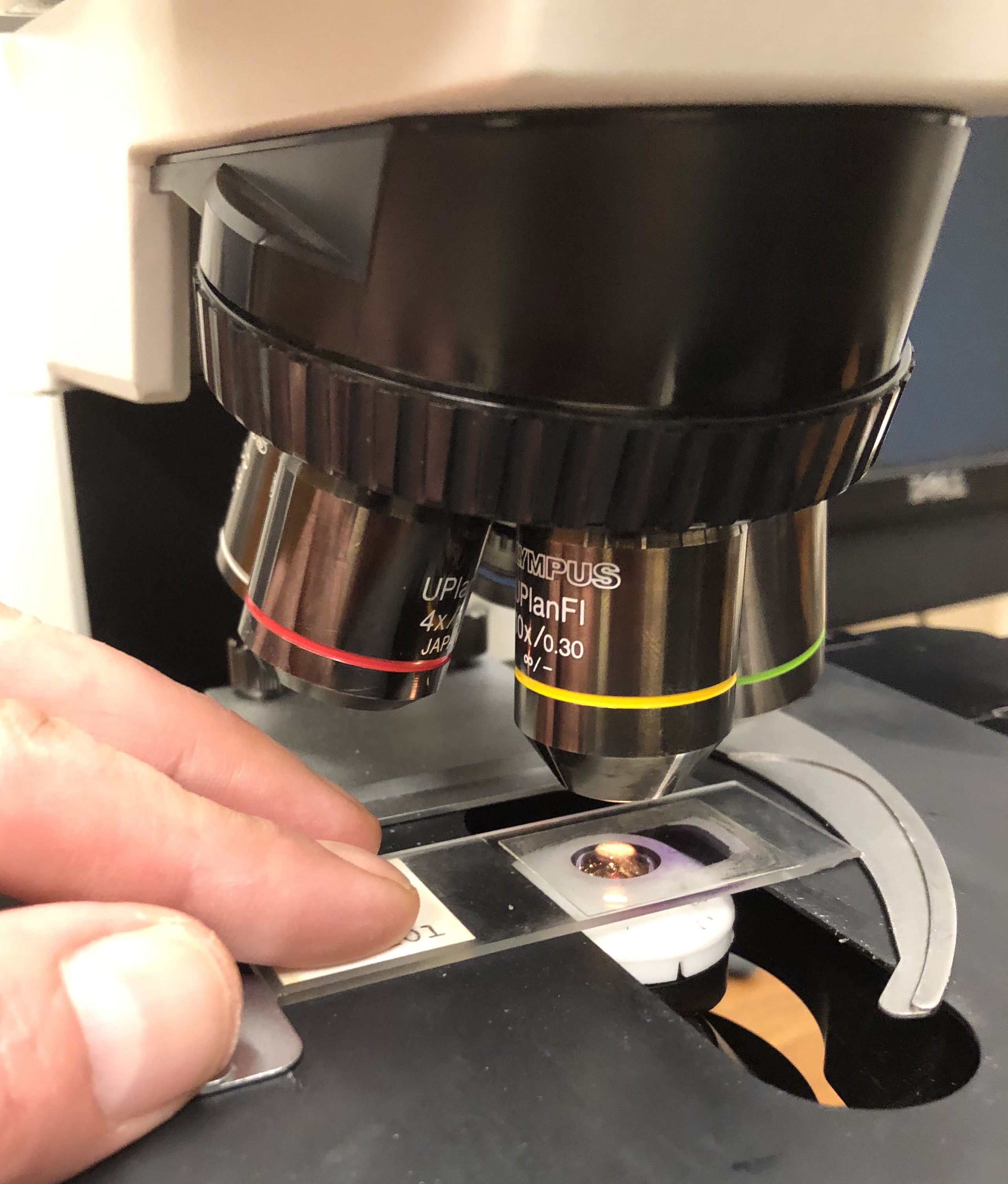
** Histology is the microscopic study of the structure, organization, and function of animal and plant tissues, providing the cellular bridge between gross anatomy and molecular biology.
**CONTENT:**
## Overview
Histology—also called **microscopic anatomy**, **microanatomy**, or **histoanatomy**—examines tissues at magnifications ranging from 40× to 1,000×, typically using light microscopes equipped with lenses that resolve details as fine as 0.2 µm. By preparing thin sections (often 5–10 µm thick) and staining them with dyes such as **hematoxylin and eosin (H&E)**, researchers can distinguish cell types, extracellular matrix components, and vascular networks that are invisible to the naked eye. Modern histology integrates **immunohistochemistry**, **fluorescence microscopy**, and **digital image analysis**, allowing scientists to map protein expression, track disease progression, and even reconstruct three‑dimensional tissue architecture from serial sections.
The discipline serves as the microscopic counterpart to **gross anatomy**, which surveys organs and systems visible without magnification. While gross anatomy tells us where a heart sits in the thorax, histology reveals the layered arrangement of **myocardial fibers**, **intercalated discs**, and **capillary networks** that enable its contractile function. In medicine, histological examination of biopsy samples remains the gold standard for diagnosing cancers, inflammatory disorders, and infectious diseases, making it indispensable for both research and clinical practice.
## History/Background
The roots of histology trace back to the invention of the compound microscope in the early 17th century. In **1665**, **Robert Hooke** published *Micrographia*, describing “cells” in cork—a term derived from the Latin *cella* (small room). However, it was not until **1805** that **Johann Heinrich Meckel** coined the term **“histology”** to denote the study of tissues. The 19th century saw rapid advances: **Camillo Golgi** introduced the **black reaction** in **1873**, allowing visualization of individual neurons, while **Santiago Ramón y Cajal** refined silver staining techniques in **1888**, laying the foundation for modern neurohistology.
The development of **paraffin embedding** in the **1890s** standardized tissue processing, and the introduction of **microtomes** capable of cutting sections as thin as 2 µm in **1900** dramatically improved image clarity. The mid‑20th century brought **immunohistochemistry** (first described in **1941** by **Albert Coons**) and **electron microscopy** (commercially available by the **1970s**), expanding the resolution limit to sub‑nanometer scales. In the 21st century, **digital pathology** and **AI‑driven image analysis** (emerging around **2015**) have begun to automate diagnostic workflows, heralding a new era of quantitative histology.
## Key Information
- **Tissue Types:** Epithelial, connective, muscle, and nervous tissues constitute the four primary histological categories, each with distinct cellular morphology and extracellular matrix composition.
- **Staining Techniques:** Beyond H&E, specialized stains include **Masson’s trichrome** (collagen), **Periodic acid‑Schiff (PAS)** (glycogen), and **Ziehl‑Neelsen** (acid‑fast bacteria).
- **Microscopy Modalities:** Light microscopy (bright‑field, phase‑contrast), fluorescence microscopy, confocal laser scanning, and **multiphoton microscopy** enable visualization from the organ level down to sub‑cellular organelles.
- **Quantitative Metrics:** Modern histology quantifies cell density (cells mm⁻³), nuclear-cytoplasmic ratio, and staining intensity using software such as **ImageJ** or **QuPath**, providing objective data for research and pathology.
- **Clinical Applications:** Histopathology diagnoses > 80 % of cancers, guides treatment decisions (e.g., HER2 status in breast cancer), and monitors transplant rejection via **C4d staining**.
## Significance
Histology underpins our understanding of how form dictates function in living systems. By revealing the micro‑architecture of organs, it informs **physiological models**, **pharmacological targeting**, and **regenerative medicine** strategies such as **organoid culture** and **3‑D bioprinting**, where tissue‑level fidelity is crucial. In evolutionary biology, comparative histology uncovers conserved cellular patterns across species, shedding light on developmental pathways. Clinically, the ability to detect microscopic abnormalities early—often before symptoms arise—has saved countless lives, exemplified by routine **Pap smear** screening that reduces cervical cancer incidence by > 70 % since its widespread adoption in the 1960s. As computational tools evolve, histology is poised to become a **big‑data discipline**, integrating genomics, proteomics, and spatial transcriptomics to construct comprehensive tissue atlases, such as the **Human Cell Atlas** launched in **2016**.
**INFOBOX:**
- Name: Histology (Microscopic Anatomy)
- Type: Biological science sub‑discipline
- Date: Established 1805 (term coined)
- Location: Global (laboratories, hospitals, research institutes)
- Known For: Detailed study of tissue structure, diagnostic pathology, and foundation for modern biomedical research
**TAGS:** histology, microscopy, tissue anatomy, pathology, immunohistochemistry, digital pathology, cell biology, biomedical research
Science
Histology
** Histology is the microscopic study of the structure, organization, and function of animal and plant tissues, providing the cellular bridge between gross anatomy and molecular biology.
**CONTENT:**
## Overview
Histology—also called **microscopic anatomy**, **microanatomy**, or **histoanatomy**—examines tissues at magnifications ranging from 40× to 1,000×, typically using light microscopes equipped with lenses that resolve details as fine as 0.2 µm. By preparing thin sections (often 5–10 µm thick) and staining them with dyes such as **hematoxylin and eosin (H&E)**, researchers can distinguish cell types, extracellular matrix components, and vascular networks that are invisible to the naked eye. Modern histology integrates **immunohistochemistry**, **fluorescence microscopy**, and **digital image analysis**, allowing scientists to map protein expression, track disease progression, and even reconstruct three‑dimensional tissue architecture from serial sections.
The discipline serves as the microscopic counterpart to **gross anatomy**, which surveys organs and systems visible without magnification. While gross anatomy tells us where a heart sits in the thorax, histology reveals the layered arrangement of **myocardial fibers**, **intercalated discs**, and **capillary networks** that enable its contractile function. In medicine, histological examination of biopsy samples remains the gold standard for diagnosing cancers, inflammatory disorders, and infectious diseases, making it indispensable for both research and clinical practice.
## History/Background
The roots of histology trace back to the invention of the compound microscope in the early 17th century. In **1665**, **Robert Hooke** published *Micrographia*, describing “cells” in cork—a term derived from the Latin *cella* (small room). However, it was not until **1805** that **Johann Heinrich Meckel** coined the term **“histology”** to denote the study of tissues. The 19th century saw rapid advances: **Camillo Golgi** introduced the **black reaction** in **1873**, allowing visualization of individual neurons, while **Santiago Ramón y Cajal** refined silver staining techniques in **1888**, laying the foundation for modern neurohistology.
The development of **paraffin embedding** in the **1890s** standardized tissue processing, and the introduction of **microtomes** capable of cutting sections as thin as 2 µm in **1900** dramatically improved image clarity. The mid‑20th century brought **immunohistochemistry** (first described in **1941** by **Albert Coons**) and **electron microscopy** (commercially available by the **1970s**), expanding the resolution limit to sub‑nanometer scales. In the 21st century, **digital pathology** and **AI‑driven image analysis** (emerging around **2015**) have begun to automate diagnostic workflows, heralding a new era of quantitative histology.
## Key Information
- **Tissue Types:** Epithelial, connective, muscle, and nervous tissues constitute the four primary histological categories, each with distinct cellular morphology and extracellular matrix composition.
- **Staining Techniques:** Beyond H&E, specialized stains include **Masson’s trichrome** (collagen), **Periodic acid‑Schiff (PAS)** (glycogen), and **Ziehl‑Neelsen** (acid‑fast bacteria).
- **Microscopy Modalities:** Light microscopy (bright‑field, phase‑contrast), fluorescence microscopy, confocal laser scanning, and **multiphoton microscopy** enable visualization from the organ level down to sub‑cellular organelles.
- **Quantitative Metrics:** Modern histology quantifies cell density (cells mm⁻³), nuclear-cytoplasmic ratio, and staining intensity using software such as **ImageJ** or **QuPath**, providing objective data for research and pathology.
- **Clinical Applications:** Histopathology diagnoses > 80 % of cancers, guides treatment decisions (e.g., HER2 status in breast cancer), and monitors transplant rejection via **C4d staining**.
## Significance
Histology underpins our understanding of how form dictates function in living systems. By revealing the micro‑architecture of organs, it informs **physiological models**, **pharmacological targeting**, and **regenerative medicine** strategies such as **organoid culture** and **3‑D bioprinting**, where tissue‑level fidelity is crucial. In evolutionary biology, comparative histology uncovers conserved cellular patterns across species, shedding light on developmental pathways. Clinically, the ability to detect microscopic abnormalities early—often before symptoms arise—has saved countless lives, exemplified by routine **Pap smear** screening that reduces cervical cancer incidence by > 70 % since its widespread adoption in the 1960s. As computational tools evolve, histology is poised to become a **big‑data discipline**, integrating genomics, proteomics, and spatial transcriptomics to construct comprehensive tissue atlases, such as the **Human Cell Atlas** launched in **2016**.
**INFOBOX:**
- Name: Histology (Microscopic Anatomy)
- Type: Biological science sub‑discipline
- Date: Established 1805 (term coined)
- Location: Global (laboratories, hospitals, research institutes)
- Known For: Detailed study of tissue structure, diagnostic pathology, and foundation for modern biomedical research
**TAGS:** histology, microscopy, tissue anatomy, pathology, immunohistochemistry, digital pathology, cell biology, biomedical research
 People
Jane Goodall
Dame Jane Goodall revolutionized primatology through her 60-year field study of wild chimpanzees, proving that humans are not the only species to make tools, show compassion, and wage war.
People
Jane Goodall
Dame Jane Goodall revolutionized primatology through her 60-year field study of wild chimpanzees, proving that humans are not the only species to make tools, show compassion, and wage war.
 People
Louis Pasteur
Louis Pasteur (1822-1895) was a French chemist and microbiologist who revolutionized medicine through his discovery of germ theory, development of pasteurization, and creation of life-saving vaccines for rabies and anthrax.
People
Louis Pasteur
Louis Pasteur (1822-1895) was a French chemist and microbiologist who revolutionized medicine through his discovery of germ theory, development of pasteurization, and creation of life-saving vaccines for rabies and anthrax.
 People
Murray Gell-Mann
Murray Gell-Mann was the American theoretical physicist who discovered the quark—the fundamental constituent of matter—and brought order to the particle zoo through his Eightfold Way classification, earning the 1969 Nobel Prize in Physics.
People
Murray Gell-Mann
Murray Gell-Mann was the American theoretical physicist who discovered the quark—the fundamental constituent of matter—and brought order to the particle zoo through his Eightfold Way classification, earning the 1969 Nobel Prize in Physics.
 People
Robert Koch
Robert Koch was the German physician who transformed medicine by proving that specific microscopic organisms cause specific diseases, earning the 1905 Nobel Prize for his isolation of the tuberculosis bacillus.
People
Robert Koch
Robert Koch was the German physician who transformed medicine by proving that specific microscopic organisms cause specific diseases, earning the 1905 Nobel Prize for his isolation of the tuberculosis bacillus.